
Found a New Lump on Your Pet? What to Record
On this page
Quick answer
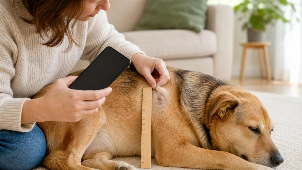
When you find a new lump on a dog or cat, note its exact location, date found, approximate size, shape, skin appearance, firmness, movement, and whether it is painful, warm, bleeding, or changing. Photograph it beside a ruler without squeezing it, then arrange a veterinary exam. Appearance and touch cannot reliably identify a lump; sampling may be needed.
Why a lump needs veterinary assessment
A lump is a localized swelling, mass, or thickened area that may arise from skin, fat, glands, infection, inflammation, fluid, injury, or abnormal tissue growth. Many causes can look similar from the outside.
Age, breed, location, texture, and growth pattern may inform a veterinarian’s assessment, but none can confirm a diagnosis alone. A soft movable mass can still need testing, and a firm mass is not automatically cancer.
How to record it safely
- Map the location. Write the body side and a fixed landmark, such as “left chest, two finger-widths behind front leg.”
- Measure consistently. Use a flexible ruler or calipers only if the pet remains relaxed. Record length and width in one unit.
- Take clear photos. Include a ruler, use the same angle and lighting, and capture the surrounding body area for orientation.
- Describe the surface. Note normal skin, hair loss, redness, ulceration, crust, discharge, or bruising.
- Observe behavior. Record licking, scratching, limping, appetite change, reduced energy, weight change, or discomfort.
- Check the rest of the body gently. Do not repeatedly press, massage, puncture, or attempt to drain any lump.
A lump map is a simple body diagram or written list used to track multiple masses by location and date. Number each one so the veterinary team can match records to the same site.
Choose the right urgency
Arrange a prompt routine appointment: any new persistent lump, an old lump that has not been assessed, or a known lump that changes size, firmness, color, movement, or symptoms.
Seek faster veterinary advice: rapid growth, pain, heat, redness, discharge, bleeding, ulceration, interference with walking, eating, swallowing, urination, defecation, or sleep; associated fever, appetite loss, weight loss, or lethargy.
Emergency care: breathing difficulty, collapse, uncontrolled bleeding, severe pain, facial or throat swelling, rapidly expanding swelling after a bite, sting, medication, or exposure, or another sudden life-threatening change.
Best approach: document once and get professional guidance. Not ideal: waiting for a mass to become large, treating it with human creams, or relying on internet photos to name it.
Appointment checklist
- Date first noticed and whether it appeared suddenly or gradually
- Body map and current measurements
- Time-stamped photos showing change
- Pain, licking, bleeding, discharge, and functional effects
- Recent wounds, injections, bites, travel, illness, or procedures
- Current medications and supplements
- History of previous masses and laboratory results
- Questions about examination, sampling, monitoring, and follow-up
A veterinarian may recommend observation, a fine-needle sample, cytology, biopsy, imaging, removal, or other testing based on the full assessment. Ask what each test can and cannot determine and when results or rechecks are expected.
Limitations and important notes
This article is general veterinary education and cannot diagnose a mass. Measuring at home can be imprecise because fur, skin movement, pet position, and pressure change the apparent borders.
Do not insert a needle, squeeze, cut, tie off, freeze, or apply caustic or human products to a lump. Doing so can cause pain, infection, bleeding, tissue damage, and delayed diagnosis. Prevent licking only with a veterinary-approved method that does not impair breathing, eating, movement, or circulation.
Sources and evidence notes
This guide reflects common veterinary practice: document location and change, assess the whole animal, and use tissue or cell sampling when indicated because palpation and photographs alone are not definitive. It does not assign a diagnosis from a visual feature.
Frequently asked questions
Can I watch a lump for a few weeks?
Ask the veterinarian rather than choosing a delay yourself. If monitoring is appropriate, obtain a specific measurement method, interval, and action threshold.
Does a movable lump mean it is harmless?
No. Mobility is one examination feature, not a diagnosis. Testing may still be recommended.
Could it be a tick?
Possibly, but do not pull an unidentified skin structure. Look closely without squeezing and contact the clinic if uncertain.
Why did a needle sample not give an answer?
Some masses shed few cells or contain mixed material. The veterinarian may recommend repeat sampling, biopsy, imaging, or removal.
Should I measure every day?
Frequent handling may irritate the area and magnify measurement noise. Use the schedule your veterinary team recommends.
Next steps
Create one labeled photo and measurement today, stop manipulating the area, and call a veterinary clinic with the location, duration, and any urgent signs. Bring the complete record so the team can assess change and choose the next diagnostic step.



 VCA Cromwell Animal Hospital4.0 (214 reviews)
 North Kenilworth Veterinary Care4.0 (586 reviews)
 St. Boni Pet Hospital4.0 (240 reviews)
 MedVet Indianapolis4.0 (938 reviews)
 Tidewater Equine Clinic & Farm Animal Services4.0 (49 reviews)
 West Riverside Veterinary Hospital4.0 (104 reviews)
 How to Prevent and Treat Heatstroke in Dogs During Summer: A Complete Guide
 The Benefits of Regular Vet Check-Ups for Your Pet's Longevity and Quality of Life
 Expert Advice on Pet Fitness for American Families
 How to Choose the Best Pet Toys for Large Dogs
 Tips for Feeding Senior Dogs with Sensitive Stomachs and Improving Digestion
 How to Safely Trim Your Dog's Nails at Home